Point
4
Intro
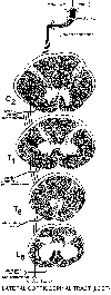 Cells in the cerebral cortex, especially the
motor cortex (area 4; precentral gyrus) possess very long
axons that descend through an extensive region of the brain
to eventually reach the spinal cord. Right before entering
the cord these corticospinal fibers cross or decussate (L.,
to make an X) and enter the LATERAL FUNICULUS where
they travel medial to the DSCT. These fibers,
which are now called the lateral (they are in the
lateral funiculus) corticospinal tract (LCST),
innervate neurons in the spinal cord along its entire
length. Once in the grey matter (where the cells are) LCST
axons synapse upon cells in the ventral horn. This is the
first synapse in a pathway over which the cerebral cortex
informs cells in the CONTRALATERAL spinal cord about a
voluntary movement that it wishes to perform. Once the
cells in the ventral horn receive this cortical information,
they directly drive the muscles via axons that pass
out the ventral root. The fastest way you can move a body
muscle voluntarily is by utilizing 2 neurons. The
first one lies in the cerebral cortex, the
second in the contralateral or opposite ventral
horn.
Cells in the cerebral cortex, especially the
motor cortex (area 4; precentral gyrus) possess very long
axons that descend through an extensive region of the brain
to eventually reach the spinal cord. Right before entering
the cord these corticospinal fibers cross or decussate (L.,
to make an X) and enter the LATERAL FUNICULUS where
they travel medial to the DSCT. These fibers,
which are now called the lateral (they are in the
lateral funiculus) corticospinal tract (LCST),
innervate neurons in the spinal cord along its entire
length. Once in the grey matter (where the cells are) LCST
axons synapse upon cells in the ventral horn. This is the
first synapse in a pathway over which the cerebral cortex
informs cells in the CONTRALATERAL spinal cord about a
voluntary movement that it wishes to perform. Once the
cells in the ventral horn receive this cortical information,
they directly drive the muscles via axons that pass
out the ventral root. The fastest way you can move a body
muscle voluntarily is by utilizing 2 neurons. The
first one lies in the cerebral cortex, the
second in the contralateral or opposite ventral
horn.
 Descending fibers in the
LCST are somatotopically organized such that the most
medially located fibers in the tract terminate before
(rostral to) the more the laterally
placed fibers.
Descending fibers in the
LCST are somatotopically organized such that the most
medially located fibers in the tract terminate before
(rostral to) the more the laterally
placed fibers.